Psychiatry Investig Search

CLOSE


Psychiatry Investig > Volume 20(11); 2023 > Article
Ahn, Lee, and Lee: Impact of Intolerance of Uncertainty on Brain Structural Changes in Panic Disorder

Abstract

Objective

This study investigated the impact of intolerance of uncertainty (IU) on structural changes in the brain and symptom severity in patients with panic disorder.

Methods

This study included 90 participants diagnosed with panic disorder. The IU Scale, Panic Disorder Severity Scale (PDSS), Beck Depression Inventory-II (BDI-II), Penn State Worry Questionnaire (PSWQ), Self-Forgiveness Scale (SFS), and Short Form 36 Health Survey (SF) were used. A voxel-wise correlation analysis was conducted to investigate the structural differences in the gray matter.

Results

As IU increased, the cortical thickness of the right lingual gyrus decreased significantly, while the gray matter volume of the right pars triangularis increased. The cortical thickness of the right lingual gyrus showed a significant negative correlation with the BDI-II score and a positive correlation with the SFS. Additionally, the gray matter volume of the right pars triangularis was positively correlated with the PDSS, PSWQ, and BDI-II scores and negatively correlated with the mental health domain of the SF.

Conclusion

According to our findings, elevated IU in participants with panic disorder was associated with cortical thinning in the lingual gyrus and increased gray matter volume in the pars triangularis. These structural alterations may also have an impact on perceived quality of life, as well as high levels of depression and anxiety.

INTRODUCTION

Panic disorder (PD) is an anxiety disorder characterized by unexpected and recurrent panic attacks [1]. In addition to panic attacks, PD has two core symptoms, maladaptive behaviors related to panic attacks and repetitive worries about them, known as anticipatory anxiety [2]. Recurrent and unpredictable panic attacks are associated with persistent anticipatory anxiety, which exacerbates the development of PD [3]. In this context, the ability to predict and anticipate situations plays an important role in the progression of PD.
Intolerance of uncertainty (IU) is a dispositional inclination to react negatively to unknown situations and events at emotional, cognitive, and behavioral levels [4]. IU was initially used to distinguish individuals with a generalized anxiety disorder from healthy individuals [5], but research has subsequently been conducted on its association with other anxiety disorders, such as social anxiety disorder [6-8], post-traumatic stress disorder [9,10], and obsessive-compulsive disorder [11,12].
Furthermore, studies on the relationship between PD and IU have also been conducted owing to these characteristics. In a previous study, the PD patient group had higher IU Scale (IUS) scores than the healthy control group [13], and the IU inhibitory factor demonstrated a significant correlation with the severity of panic symptoms [14]. Anxiety plays a role in altering attentional processes when coping with threatening stimuli [15]. Anxious individuals tend to interpret ambiguous and neutral stimuli as threatening, which is known as an “interpretation bias.” [16] Patients with PD have a negative cognitive tendency to interpret ambiguous physiological sensations, which can be a risk factor for disease progression [17].
The neural pathways associated with IU have been studied to understand these cognitive processes. Numerous studies have been conducted to enhance understanding of the neurological basis of IU. Many functional magnetic resonance imaging (MRI) studies have demonstrated a positive correlation between anterior insula activity and IU [18-21]. Additionally, associations between anterior cingulate cortex and amygdala activity have also been observed [22,23]. It is widely accepted that PD involves the disruption of the fear circuit centered around the amygdala [24,25]. The interconnectedness between the amygdala, insula, and anterior cingulate, with the amygdala at its core, integrates sensory and bodily inputs and their interpretation of these inputs.26,27 Hypervigilance toward anticipated threats and responses is associated with amygdala overactivity [28], which is richly connected to the orbitofrontal cortex, ventral striatum, and sensory cortex [29]. The alteration of these connections makes it difficult to distinguish between safe and threatening situations, and this uncertainty is thought to be associated with anxiety [30]. In addition, there are studies that have shown associations between IU and brain structural changes. Hilbert et al. [31] reported a positive correlation between IU and gray matter volume (GMV) in the right temporal gyri in individuals with generalized anxiety disorder. Additionally, Kim et al. [32] found a positive correlation between striatal volume and IU in healthy participants.
However, little research has been conducted on structural changes in the brain associated with IU in patients with PD, and even less is known about how these structural changes affect actual functioning. Therefore, this study aimed to investigate the brain regions and structural associations of IU in patients with PD, as well as the impact of these structural alterations on clinical outcomes.

METHODS

Participants

Ninety participants with PD, aged between 18 and 66 years, were included in the current study. The participants were recruited from the Department of Psychiatry at CHA Bundang Medical Center between December 2013 and June 2020. All participants were right-handed Koreans and underwent MRI. Qualified psychiatrists diagnosed PD using the Structured Clinical Interview for the Diagnostic and Statistical Manual of Mental Disorders, Fifth Edition [2].
The exclusion criteria were as follows: a primary diagnosis of schizophrenia, anxiety disorders other than PD, major depressive disorder, substance use disorder, or mental retardation. Participants who were unable to undergo brain MRI were excluded.
The study protocols followed the Good Clinical Practice guidelines and were approved by the CHA Bundang Medical Center’s Institutional Review Board (no. 2019-05-030, 2021-05-055). Written informed consent was obtained from all participants after they were given a thorough explanation of the study.

Assessments

To evaluate negative reactions to uncertainty, the Korean version of the IUS-Short Form (IUS-12) was used. The original scale, the IUS, was created by Freeston in 1994. Later, Carleton et al. [33] developed and validated a shortened 12-item, two-factor version of the scale in 2007. The scale is measured on a 4-point Likert scale and consists of 12 items that evaluate responses to uncertainty, ambiguity, and the future. Cronbach’s α for the Korean version of the IUS-12 was 0.94 [34].
To assess the severity of the participants’ baseline anxiety and depression, the Panic Disorder Severity Scale (PDSS) [35], Beck Depression Inventory-II (BDI-II) [36], Beck Anxiety Inventory (BAI) [37], and Penn State Worry Questionnaire (PSWQ) were used [38].
The PDSS consists of items that assess PD and seven panic-associated symptoms. The Korean version of the PDSS has demonstrated strong reliability, validity, and internal consistency (Cronbach’s α=0.89) [39]. The PSWQ is a tool used to measure pathological levels of anxiety and worry. The Korean version of the PSWQ has shown good internal consistency (Cronbach’s α=0.90) and factorial validity [40].
The BDI is one of the most commonly used screening tests for depression, evaluating depressive symptoms over the past two weeks. The Korean version of the BDI has shown high internal consistency (Cronbach’s α=0.94) and proper validity [41]. The BAI is a tool used to measure anxiety. The Korean version of the BAI has demonstrated favorable internal consistency (Cronbach’s α=0.90) and validity [42].
The Self-Forgiveness Scale (SFS) was used to evaluate dispositional self-forgiveness. The SFS comprises three subscales: “acceptance and improvement,” “transferring responsibility,” and “negative affect, thoughts, and behavior.” The Korean version of the SFS has demonstrated adequate validity and high internal consistency (Cronbach’s α=0.87) [43].
Additionally, to measure perceived life satisfaction, the Korean version of the Short Form 36 Health Survey (SF) was used. The SF measures health-related quality of life and is categorized into eight domains: physical functioning, social functioning, physical role, emotional role, mental health, vitality, bodily pain, and general health [31]. The Korean version of the SF has shown satisfactory content and discriminant validity, with Cronbach’s α ranging from 0.71 to 0.90 [44].

Neuroimaging data acquisition

All subjects underwent MRI using the 3T GE Signa HDxt scanner (GE Healthcare, Milwaukee, WI, USA) with an eight-channel phase-array head coil. The settings for acquiring T1-weighted fast spoiled gradient recalled echo images in the three dimensions were as follows: 6.3 ms repetition time; 2.1 ms echo time; 12° flip angle; 1 mm slice thickness; 25.6 cm field of view; 256×256 matrix; and 1×1×1 mm3 isotropic voxel size.
To analyze GMV and cortical thickness (CT), FreeSurfer v7.1.0 (http://surfer.nmr.mgh.harvard.edu) was used. Image processing was performed as follows: motion correction, intensity normalization, skull stripping, white matter segmentation, gray/white matter border tesselation, automated topology correction, and surface deformation. Previous studies have provided descriptions of the specificities of these methods in detail [45-47]. The cortex was auto-parcellated into 34 separate gyral regions per hemisphere utilizing gyral and sulcal architecture using the Desikan-Killiany atlas [48-50].

Statistical analysis

Age, sex, and the total intracranial volume (ICV) were included as covariates in the multiple regression models. We conducted the Monte-Carlo simulation to correct for multiple comparisons in cluster-wise correction.
In addition, to identify the significance of the CT in the brain correlates of the IUS and its associations with the psychological characteristics of patients with PD, an exploratory correlation analysis was performed. Pearson’s correlation analysis was performed using the IBM Statistical Package for the Social Sciences Windows software version 27 (IBM Corp., Armonk, NY, USA). A p-value of 0.05 or less was regarded as statistically significant.

RESULTS

Sociodemographics

Of the 90 participants with PD, 45 (50.0%) were male, and 45 (50.0%) were female (Table 1). The mean age of the participants was 37.92 years (standard deviation [SD]=12.57), and the mean number of years they spent in school was 14.61 (SD=2.29). A summary of the sociodemographic characteristics is presented in Table 1.

Association between IUS and gray matter structural alteration

In PD, there was a significant negative correlation between the total IUS scores and the CT in the right lingual gyrus (peak voxel of MNI: X=15.4, Y=-74.5, Z=-6.5; Monte Carlo simulation correction, cluster-wise p<0.05) (Figure 1A). There was also a significant positive correlation between the total IUS scores and GMV in the right pars triangularis (peak voxel of MNI: X=39.1, Y=33.8, Z=9.0; Monte Carlo simulation correction, cluster-wise p<0.05) (Figure 1B). After controlling for variables including age, sex, and ICV, statistical significance was maintained.

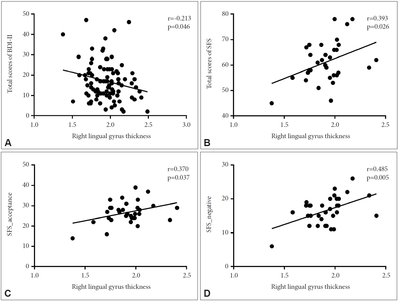
Correlation between the mean CT in the right lingual gyrus and clinical scales of PD

In participants with PD, exploratory correlation analysis showed significant negative associations between the CT in the right lingual gyrus and the total BDI-II scores. In addition, strong positive correlations were found between the CT in the right lingual gyrus and the total scores of the SFS and SFS subscales (negative emotional status and acceptance) (Figure 2).

Correlation between the GMV in the right pars triangularis and clinical scales of PD

In addition, there was a significant negative correlation between the GMV in the right pars triangularis and the mental health domain of the SF. There was a positive correlation between the GMV and the baseline total scores on the PDSS, PSWQ, and BDI-II, and the transferring responsibility subscales of the SFS (Figure 3).

DISCUSSION

This study investigated the structural brain alterations associated with IU and the associated psychological characteristics in patients with PD. Although the regions identified in previous structural-based studies did not show significant differences in the present study, it is important to note that this study employed a whole brain analysis approach specifically targeting patients with PD. Considerable amount of research has provided evidence that IU is a transdiagnostic dispositional risk factor for the development and maintenance of clinically significant anxiety [19,31,51]. Growing evidence indicates that IU is associated with a broad range of emotional disorders and other cognitive vulnerability factors [52,53], highlighting the theoretical and therapeutic importance of IU. Recent researchers argue that disorder-specific IU is more strongly related to symptoms of disorder than general IU trait [54]. This difference sets the present study apart from previous studies and provides a unique perspective on the neural correlates of PD. In patients with PD, high IU was significantly correlated with lingual gyrus thinning and increased GMV in the pars triangularis. Furthermore, the CT of the lingual gyrus was negatively correlated with subjective depression, whereas self-forgiveness and the tendency to accept mistakes and avoid negative aspects were positively correlated. The GMV in the pars triangularis was positively correlated with excessive anxiety levels, depression, and a sense of personal responsibility for mistakes but negatively correlated with mental health satisfaction.
In this study, a higher IUS score in patients with PD was associated with decreased CT in the lingual gyrus. Due to their sensitivity to environmental changes, patients with PD tend to perceive even secure situations as threatening [55], which may be a precipitating factor in PD [56,57]. According to previous studies, as part of the visual cortex, the lingual gyrus is associated with the amygdala, modulates spatial attention and fear responses [58], and is hyperactive in response to uncertain stimuli [59]. Therefore, errors in the interpretation of sensory information in the visual cortex, including the lingual gyrus, may play a significant role in the hypervigilance of neutral stimuli.
This study also found that high IU levels in patients with PD were associated with greater GMV in the pars triangularis. These findings align with previous research that has shown an association between IU and increased volume in the superior temporal pole, which is involved in affective face processing [31]. Part of the ventrolateral prefrontal cortex, the pars triangularis, is associated with affective regulation and fear conditioning in patients with PD [60,61]. The right inferior frontal gyrus is also involved in detecting behaviorally relevant stimuli that are salient or unexpected [62]. Previous research by Weilnhammer et al. [63] demonstrated that the inferior frontal gyrus contributes to the interpretation of ambiguous sensory stimuli via feedback processes with the visual cortex [63-66]. Based on these findings, structural alterations in the pars triangularis and the lingual gyrus may result in erroneous interpretations of uncertain stimuli and anxiety [66].
Thinning of the lingual gyrus, which is associated with high IUS scores, is associated with severe depressive symptoms, whereas an increase in the volume of the pars triangularis is positively correlated with PDSS, PSWQ, and BDI-II scores. This is consistent with a meta-analysis conducted by Arnone et al. [67] that demonstrated a significant decrease in the gray matter density of the lingual gyrus in patients with depression [67,68]. In addition, previous studies on patients with PD have demonstrated a positive correlation between the pars triangularis CT and the severity of panic symptoms, supporting the findings of this study [69].
Thinning of the lingual gyrus and increased size of the pars triangularis appeared to diminish life satisfaction. Numerous studies have demonstrated that IU is associated with coping styles, resilience, forgiveness, self-compassion, and lower quality of life, as higher IU levels tend to be associated with lower life satisfaction [70-73]. This is thought to be due to a genetically susceptible brain structure that increases “interpretation bias” toward neutral stimuli, making individuals hypervigilant, which exacerbates anxiety and depressive symptoms and results in a decline in quality of life [16].
This study has several limitations. First, because this study was limited to patients with PD, generalizing the results to the general population may be challenging. Nevertheless, this study provides a convincing explanation for the neural basis of IU in PD. Second, because this was a cross-sectional study, it could be difficult to establish causality between the results. Therefore, further research may be required to investigate the changes in IUS over time and the accompanying structural alterations in the brain.
In conclusion, the results of our study indicate that elevated IU in patients with PD is associated with cortical thinning in the lingual gyrus and increased volume in the pars triangularis, which may be related to high levels of individual depression and anxiety as well as decreased quality of life. This indicates that the lingual gyrus and pars triangularis may contribute to interpretation bias, resulting in hypersensitivity to neutral stimuli and increased IU in uncertain situations. These findings enhance the neurological comprehension of PD-specific anticipatory anxiety and IU.

Notes

Availability of Data and Material

The datasets generated or analyzed during the study are not publicly available due to ethical restrictions for the protection of personal and medical information of participants but are available from the corresponding author on reasonable request with the approval of the Institutional Review Board of CHA Bundang Medical Center.

Conflicts of Interest

Kang Soo Lee, a contributing editor of the Psychiatry Investigation, was not involved in the editorial evaluation or decision to publish this article. All remaining authors have declared no conflicts of interest.

Author Contributions

Conceptualization: Sungjun Ahn, Sang-Hyuk Lee. Data curation: all authors. Formal analysis: Sungjun Ahn. Funding acquisition: Sang-Hyuk Lee. Investigation: Sungjun Ahn, Sang-Hyuk Lee. Methodology: Sungjun Ahn, Sang-Hyuk Lee. Project administration: Kang Soo Lee, Sang-Hyuk Lee. Resources: Kang Soo Lee, Sang-Hyuk Lee. Software: Sungjun Ahn. Supervision: Kang Soo Lee. Validation: Kang Soo Lee. Visualization: Sungjun Ahn, Kang Soo Lee. Writing—original draft: Sungjun Ahn. Writing—review & editing: Sang-Hyuk Lee, Kang Soo Lee.

Funding Statement

This study was supported by the Basic Science Research Program through the National Research Foundation of Korea, funded by the Ministry of Science and ICT (grant number NRF-2019M3C7A1032262 and grant number NRF-2021M3E5D9025026). This study was also funded in part by the Healthcare AI Convergence Research & Development Program through the National IT Industry Promotion Agency of Korea (NIPA), funded by the Ministry of Science and ICT (grant number S0102-23-1008). Both sets of funding were secured by S.H. Lee.

Figure 1.
Association between IUS and gray matter structural alteration. A: Cluster with significant negative correlations between the intolerance of uncertainty total scores and the cortical thickness of the lingual gyrus in the right hemisphere (peak voxel of MNI: X=15.4, Y= -74.5, Z=-6.5) in individuals with panic disorder (cluster p<0.05). B: Cluster with significant positive correlations between the intolerance of uncertainty total scores and the gray matter volume of the pars triangularis in the right hemisphere (peak voxel of MNI: X=39.1, Y=33.8, Z= 9.0) in individuals with panic disorder (cluster p<0.05). IUS, Intolerance of Uncertainty Scale.
pi-2023-0181f1.jpg
Figure 2.
Pearson’s correlations between the cortical thickness of the right lingual gyrus and the total scores of the Beck Depression Inventory- II (BDI-II) (A), total scores of the Self-Forgiveness Scale (SFS) (B), acceptance and improvement subscales of the SFS (C), and negative affect (D), thought and behavior subscales of the SFS in patients with pain disorder.
pi-2023-0181f2.jpg
Figure 3.
Pearson’s correlations between the gray matter volume of the right pars triangularis and total scores of the Panic Disorder Severity Scale (PDSS) (A), total scores of the Penn State Worry Questionnaire (PSWQ) (B), total scores of the Beck Depression Inventory-II (BDI-II) (C), scores of transferring of responsibility on the Self-Forgiveness Scale (SFS) (D), and mental health domain scores on the Short Form 36 Health Survey (SF) (E).
pi-2023-0181f3.jpg
Table 1.
Demographic and clinical characteristics of study participants
Characteristics Value (N=90)
Sex
 Male 45 (50.0)
 Female 45 (50.0)
Age (yr) 37.92±12.57
Education (yr) 14.61±2.29
Intracranial volume (mL) 1494.54±153.91
Age of symptom onset (yr) (N=82) 33.26±12.11
Duration of illness (mo) (N=69) 46.45±79.96
Family history of anxiety disorder 24 (26.7)
Baseline IUS 31.60±6.81
Baseline PDSS 13.08±6.55
Baseline BDI-II (N=88) 17.08±9.67
Baseline PSWQ (N=87) 53.15±12.52
Baseline BAI (N=85) 23.69 ±14.82
Baseline SFS (N=32) 61.25±8.06
 Acceptance and improvement 26.72 ±5.31
 Transferring of responsibility 17.97±3.66
 Negative affect, thought, and behavior 16.56±4.13
Baseline SF (N=81) 1844.75±454.96
 Physical functioning 72.61±20.34
 Social functioning 58.10±19.70
 Role physical 28.41±37.75
 Role emotional 27.20±37.54
 Mental health 50.50±8.05
 Vitality 49.24±11.34
 Bodily pain 50.67±30.14
 General health 42.27±16.57

Values are presented as mean±standard deviation or number (%).

IUS, Intolerance of Uncertainty Scale; PDSS, Panic Disorder Severity Scale; BDI-II, Beck Depression Inventory-II; PSWQ, Penn State Worry Questionnaire; BAI, Beck Anxiety Inventory; SFS, Self-Forgiveness Scale; SF, Short-Form 36-Item Health Survey

REFERENCES

1. Klein DF, Ross DC, Cohen P. Panic and avoidance in agoraphobia. Application of path analysis to treatment studies. Arch Gen Psychiatry 1987;44:377-385.
crossref pmid
2. American Psychiatric Association. Diagnostic and Statistical Manual of Mental Disorders (5th ed). Arlington, VA: American Psychiatric Association; 2013.

3. Başoğlu M, Marks IM, Kiliç C, Swinson RP, Noshirvani H, Kuch K, et al. Relationship of panic, anticipatory anxiety, agoraphobia and global improvement in panic disorder with agoraphobia treated with alprazolam and exposure. Br J Psychiatry 1994;164:647-652.
crossref pmid
4. Carleton RN, Sharpe D, Asmundson GJ. Anxiety sensitivity and intolerance of uncertainty: requisites of the fundamental fears? Behav Res Ther 2007;45:2307-2316.
crossref pmid
5. Freeston MH, Rhéaume J, Letarte H, Dugas MJ, Ladouceur R. Why do people worry? Pers Individ Differ 1994;17:791-802.
crossref
6. Boelen PA, Reijntjes A. Intolerance of uncertainty and social anxiety. J Anxiety Disord 2009;23:130-135.
crossref pmid
7. Carleton RN, Collimore KC, Asmundson GJ. “It’s not just the judgement--It’s that I don’t know”: intolerance of uncertainty as a predictor of social anxiety. J Anxiety Disord 2010;24:189-195.
pmid
8. Counsell A, Furtado M, Iorio C, Anand L, Canzonieri A, Fine A, et al. Intolerance of uncertainty, social anxiety, and generalized anxiety: differences by diagnosis and symptoms. Psychiatry Res 2017;252:63-69.
crossref pmid
9. Fetzner MG, Horswill SC, Boelen PA, Carleton RN. Intolerance of uncertainty and PTSD symptoms: exploring the construct relationship in a community sample with a heterogeneous trauma history. Cogn Ther Res 2013;37:725-734.
crossref pdf
10. Oglesby ME, Boffa JW, Short NA, Raines AM, Schmidt NB. Intolerance of uncertainty as a predictor of post-traumatic stress symptoms following a traumatic event. J Anxiety Disord 2016;41:82-87.
crossref pmid
11. Tolin DF, Abramowitz JS, Brigidi BD, Foa EB. Intolerance of uncertainty in obsessive-compulsive disorder. J Anxiety Disord 2003;17:233-242.
crossref pmid
12. Sarawgi S, Oglesby ME, Cougle JR. Intolerance of uncertainty and obsessive-compulsive symptom expression. J Behav Ther Exp Psychiatry 2013;44:456-462.
crossref pmid
13. Carleton RN, Mulvogue MK, Thibodeau MA, McCabe RE, Antony MM, Asmundson GJ. Increasingly certain about uncertainty: intolerance of uncertainty across anxiety and depression. J Anxiety Disord 2012;26:468-479.
crossref pmid
14. Carleton RN, Duranceau S, Freeston MH, Boelen PA, McCabe RE, Antony MM. “But it might be a heart attack”: intolerance of uncertainty and panic disorder symptoms. J Anxiety Disord 2014;28:463-470.
crossref pmid
15. Bar-Haim Y, Lamy D, Pergamin L, Bakermans-Kranenburg MJ, van IJzendoorn MH. Threat-related attentional bias in anxious and nonanxious individuals: a meta-analytic study. Psychol Bull 2007;133:1-24.
crossref pmid
16. Hazlett-Stevens H, Borkovec TD. Interpretive cues and ambiguity in generalized anxiety disorder. Behav Res Ther 2004;42:881-892.
crossref pmid
17. Richards JC, Austin DW, Alvarenga ME. Interpretation of ambiguous interoceptive stimuli in panic disorder and nonclinical panic. Cogn Ther Res 2001;25:235-246.

18. Gorka SM, Nelson BD, Phan KL, Shankman SA. Intolerance of uncertainty and insula activation during uncertain reward. Cogn Affect Behav Neurosci 2016;16:929-939.
crossref pmid pdf
19. Oathes DJ, Hilt LM, Nitschke JB. Affective neural responses modulated by serotonin transporter genotype in clinical anxiety and depression. PLoS One 2015;10:e0115820.
crossref pmid pmc
20. Shankman SA, Gorka SM, Nelson BD, Fitzgerald DA, Phan KL, O’Daly O. Anterior insula responds to temporally unpredictable aversiveness: an fMRI study. Neuroreport 2014;25:596-600.
pmid pmc
21. Simmons A, Matthews SC, Paulus MP, Stein MB. Intolerance of uncertainty correlates with insula activation during affective ambiguity. Neurosci Lett 2008;430:92-97.
crossref pmid
22. Krain AL, Gotimer K, Hefton S, Ernst M, Castellanos FX, Pine DS, et al. A functional magnetic resonance imaging investigation of uncertainty in adolescents with anxiety disorders. Biol Psychiatry 2008;63:563-568.
crossref pmid
23. Schienle A, Köchel A, Ebner F, Reishofer G, Schäfer A. Neural correlates of intolerance of uncertainty. Neurosci Lett 2010;479:272-276.
crossref pmid
24. Gorman JM, Kent JM, Sullivan GM, Coplan JD. Neuroanatomical hypothesis of panic disorder, revised. Am J Psychiatry 2000;157:493-505.
crossref pmid
25. Bang M, Park YW, Eom J, Ahn SS, Kim J, Lee SK, et al. An interpretable radiomics model for the diagnosis of panic disorder with or without agoraphobia using magnetic resonance imaging. J Affect Disord 2022;305:47-54.
crossref pmid
26. Seeley WW, Menon V, Schatzberg AF, Keller J, Glover GH, Kenna H, et al. Dissociable intrinsic connectivity networks for salience processing and executive control. J Neurosci 2007;27:2349-2356.
crossref pmid pmc
27. Dresler T, Guhn A, Tupak SV, Ehlis AC, Herrmann MJ, Fallgatter AJ, et al. Revise the revised? New dimensions of the neuroanatomical hypothesis of panic disorder. J Neural Transm (Vienna) 2013;120:3-29.
crossref pmid pdf
28. Sakai Y, Kumano H, Nishikawa M, Sakano Y, Kaiya H, Imabayashi E, et al. Cerebral glucose metabolism associated with a fear network in panic disorder. Neuroreport 2005;16:927-931.
crossref pmid
29. Haber SN, Knutson B. The reward circuit: linking primate anatomy and human imaging. Neuropsychopharmacology 2010;35:4-26.
crossref pmid pdf
30. Grillon C. Associative learning deficits increase symptoms of anxiety in humans. Biol Psychiatry 2002;51:851-858.
crossref pmid
31. Hilbert K, Pine DS, Muehlhan M, Lueken U, Steudte-Schmiedgen S, Beesdo-Baum K. Gray and white matter volume abnormalities in generalized anxiety disorder by categorical and dimensional characterization. Psychiatry Res 2015;234:314-320.
crossref pmid pmc
32. Kim MJ, Shin J, Taylor JM, Mattek AM, Chavez SJ, Whalen PJ. Intolerance of uncertainty predicts increased striatal volume. Emotion 2017;17:895-899.
crossref pmid pmc
33. Carleton RN, Norton MA, Asmundson GJ. Fearing the unknown: a short version of the Intolerance of Uncertainty Scale. J Anxiety Disord 2007;21:105-117.
crossref pmid
34. Kim SH. The relationship of fear of negative and positive evaluation, intolerance of uncertainty, and social anxiety [dissertation]. Seoul, Ewha Womans University. 2010;p.26.

35. Shear MK, Brown TA, Barlow DH, Money R, Sholomskas DE, Woods SW, et al. Multicenter collaborative panic disorder severity scale. Am J Psychiatry 1997;154:1571-1575.
crossref pmid
36. Beck AT, Steer RA, Brown GK. Manual for the Beck Depression Inventory-II. San Antonio, TX: Psychological Corporation; 1996.

37. Beck AT, Epstein N, Brown G, Steer RA. An inventory for measuring clinical anxiety: psychometric properties. J Consult Clin Psychol 1988;56:893-897.
crossref pmid
38. Brown TA, Antony MM, Barlow DH. Psychometric properties of the Penn State Worry Questionnaire in a clinical anxiety disorders sample. Behav Res Ther 1992;30:33-37.
crossref pmid
39. Kim JB. Development of the Korean version of the panic disorder severity scale. Korean J Psychopathol 2001;10:140-151.

40. Jeon JW, Kim D, Kim E, Roh S. Exploratory and confirmatory factor analysis of the Korean version of the Penn State Worry Questionnaire. Anxiety Mood 2017;13:86-92.

41. Lim SY, Lee EJ, Jeong SW, Kim HC, Jeong CH, Jeon TY, et al. The validation study of Beck Depression Scale 2 in Korean version. Anxiety Mood 2011;7:48-53.

42. Lee HK, Lee EH, Hwang ST, Hong SH, Kim JH. Psychometric properties of the Beck Anxiety Inventory in the community-dwelling sample of Korean adults. Kor J Clin Psychol 2016;35:822-830.
crossref
43. Kim BJ, Lee D, Kim JG, Kim YS, Hwang JE. Development and validation of the self-forgiveness scale. Korean J Couns 2013;14:973-993.
crossref
44. Nam BH, Lee SU. Testing the validity of the Korean SF-36 health survey. J Korean Info Stat 2003;28:3-25.

45. Dale AM, Fischl B, Sereno MI. Cortical surface-based analysis. I. Segmentation and surface reconstruction. Neuroimage 1999;9:179-194.
crossref pmid
46. Fischl B, Dale AM. Measuring the thickness of the human cerebral cortex from magnetic resonance images. Proc Natl Acad Sci U S A 2000;97:11050-11055.
crossref pmid pmc
47. Fischl B, Sereno MI, Dale AM. Cortical surface-based analysis. II: inflation, flattening, and a surface-based coordinate system. Neuroimage 1999;9:195-207.
crossref pmid
48. Hagler DJ Jr, Saygin AP, Sereno MI. Smoothing and cluster thresholding for cortical surface-based group analysis of fMRI data. Neuroimage 2006;33:1093-1103.
crossref pmid
49. Desikan RS, Ségonne F, Fischl B, Quinn BT, Dickerson BC, Blacker D, et al. An automated labeling system for subdividing the human cerebral cortex on MRI scans into gyral based regions of interest. Neuroimage 2006;31:968-980.
crossref pmid
50. Fischl B, van der Kouwe A, Destrieux C, Halgren E, Ségonne F, Salat DH, et al. Automatically parcellating the human cerebral cortex. Cereb Cortex 2004;14:11-22.
crossref pmid
51. McFadden KL, Tregellas JR, Shott ME, Frank GK. Reduced salience and default mode network activity in women with anorexia nervosa. J Psychiatry Neurosci 2014;39:178-188.
crossref pmid pmc
52. Morriss J, Zuj DV, Mertens G. The role of intolerance of uncertainty in classical threat conditioning: recent developments and directions for future research. Int J Psychophysiol 2021;166:116-126.
crossref pmid
53. McEvoy PM, Mahoney AE. To be sure, to be sure: intolerance of uncertainty mediates symptoms of various anxiety disorders and depression. Behav Ther 2012;43:533-545.
crossref pmid
54. Wever M, Smeets P, Sternheim L. Neural correlates of intolerance of uncertainty in clinical disorders. J Neuropsychiatry Clin Neurosci 2015;27:345-353.
crossref pmid
55. Gorka SM, Lieberman L, Nelson BD, Sarapas C, Shankman SA. Aversive responding to safety signals in panic disorder: the moderating role of intolerance of uncertainty. J Anxiety Disord 2014;28:731-736.
crossref pmid pmc
56. Lieberman L, Gorka SM, Sarapas C, Shankman SA. Cognitive flexibility mediates the relation between intolerance of uncertainty and safety signal responding in those with panic disorder. Cogn Emot 2016;30:1495-1503.
crossref pmid
57. Craske MG, Rauch SL, Ursano R, Prenoveau J, Pine DS, Zinbarg RE. What is an anxiety disorder? Depress Anxiety 2009;26:1066-1085.
crossref pmid
58. Carlson JM, Reinke KS, Habib R. A left amygdala mediated network for rapid orienting to masked fearful faces. Neuropsychologia 2009;47:1386-1389.
crossref pmid
59. Zhang M, Ma C, Luo Y, Li J, Li Q, Liu Y, et al. Neural basis of uncertain cue processing in trait anxiety. Sci Rep 2016;6:21298
crossref pmid pmc pdf
60. Kircher T, Arolt V, Jansen A, Pyka M, Reinhardt I, Kellermann T, et al. Effect of cognitive-behavioral therapy on neural correlates of fear conditioning in panic disorder. Biol Psychiatry 2013;73:93-101.
crossref pmid
61. Lopes FL, Faria CGF, Dias GP, Mallmann MB, Mendes V, Horato N, et al. Neural correlates of negative and disease-specific emotional stimuli in panic disorder: a functional magnetic resonance imaging study. Braz J Psychiatry 2021;43:605-612.
crossref pmid pmc
62. Corbetta M, Shulman GL. Control of goal-directed and stimulus-driven attention in the brain. Nat Rev Neurosci 2002;3:201-215.
crossref pmid pdf
63. Weilnhammer V, Fritsch M, Chikermane M, Eckert AL, Kanthak K, Stuke H, et al. An active role of inferior frontal cortex in conscious experience. Current Biology 2021;31:2868-2880.
crossref pmid
64. Sterzer P, Kleinschmidt A, Rees G. The neural bases of multistable perception. Trends Cogn Sci 2009;13:310-318.
crossref pmid
65. Brascamp J, Sterzer P, Blake R, Knapen T. Multistable perception and the role of the frontoparietal cortex in perceptual inference. Annu Rev Psychol 2018;69:77-103.
crossref pmid pdf
66. Wittmann A, Schlagenhauf F, John T, Guhn A, Rehbein H, Siegmund A, et al. A new paradigm (Westphal-Paradigm) to study the neural correlates of panic disorder with agoraphobia. Eur Arch Psychiatry Clin Neurosci 2011;261:185-194.
crossref pmid pdf
67. Arnone D, Job D, Selvaraj S, Abe O, Amico F, Cheng Y, et al. Computational meta-analysis of statistical parametric maps in major depression. Hum Brain Mapp 2016;37:1393-1404.
crossref pmid pmc
68. Couvy-Duchesne B, Strike LT, de Zubicaray GI, McMahon KL, Thompson PM, Hickie IB, et al. Lingual gyrus surface area is associated with anxiety-depression severity in young adults: a genetic clustering approach. eNeuro 2018;5:ENEURO.0153-17.2017.
crossref
69. Kang EK, Lee KS, Lee SH. Reduced cortical thickness in the temporal pole, insula, and pars triangularis in patients with panic disorder. Yonsei Med J 2017;58:1018-1024.
crossref pmid pmc pdf
70. GEÇGİN FM, SAHRANÇ Ü. [The relationships between intolerance of uncertainty and psychological well-being]. Sakarya Univ J Educ 2017;7:739-756. Turkish.

71. Wang T, Jiang L, Li T, Zhang X, Xiao S. The relationship between intolerance of uncertainty, coping style, resilience, and anxiety during the COVID-19 relapse in freshmen: a moderated mediation model. Front Psychiatry 2023;14:1136084
crossref pmid pmc
72. Akgün Şahin Z, Deniz M, Akça D, Uymaz Aras G, Doğan S. The relationship between intolerance of uncertainty and spiritual well-being levels of COVID-19 among elderly: a cross sectional and correlational study. OMEGA (Westport) 2023;Mar 9 [Epub]. https://doi.org/10.1177/00302228231163208.
crossref
73. Koç M, Iskender M, Çolak TS, Düşünceli B. [Investigation of the effect of intolerance of uncertainty and the effect of anger control on the relationship between forgiveness and psychological well-being through structural equation modelling]. Sakarya Univ J Educ 2016;6:201-209. Turkish.


ABOUT
AUTHOR INFORMATION
ARTICLE CATEGORY

Browse all articles >

BROWSE ARTICLES
Editorial Office
#522, 27, Seochojungang-ro 24-gil, Seocho-gu, Seoul 06601, Korea
Tel: +82-2-717-0892    E-mail: psychiatryinvest@gmail.com                

Copyright © 2024 by Korean Neuropsychiatric Association.

Developed in M2PI

Close layer
prev next