Decreased Prolidase Activity in Patients with Posttraumatic Stress Disorder
Article information
Abstract
Objective
Many neurochemical systems have been implicated in the development of Posttraumatic Stress Disorder (PTSD). The prolidase enzyme is a cytosolic exopeptidase that detaches proline or hydroxyproline from the carboxyl terminal position of dipeptides. Prolidase has important biological effects, and to date, its role in the etiology of PTSD has not been studied. In the present study, we aimed to evaluate prolidase activity in patients with PTSD.
Methods
The study group consisted of patients who were diagnosed with PTSD after the earthquake that occurred in the province of Van in Turkey in 2011 (n=25); the first control group consisted of patients who experienced the earthquake but did not show PTSD symptoms (n=26) and the second control group consisted of patients who have never been exposed to a traumatic event (n=25). Prolidase activities in the patients and the control groups were determined by the ELISA method using commercial kits.
Results
Prolidase activity in the patient group was significantly lower when compared to the control groups. Prolidase activity was also significantly lower in the traumatized healthy subjects compared to the other healthy group (p<0.01).
Conclusion
The findings of the present study suggest that the decrease in prolidase activity may have neuroprotective effects in patients with PTSD.
INTRODUCTION
Posttraumatic stress disorder (PTSD) is a mental health problem that results after hearing about, witnessing, or experiencing a traumatic event.1 The recurring flashbacks, avoidance and numbing, and hyper-arousal are the common symptoms of PTSD. Interestingly, traumatizing events can vary and often include incidents such as car accidents, rape, war, or natural disasters all of which may threaten the life of individuals.234 In addition, PTSD is frequently encountered after earthquakes.56 In population-based studies performed after earthquakes, the rate of PTSD was reported to vary between 3.0% and 87.0%.789
The number of neurobiological studies on PTSD has increased in recent years. More than one neurochemical system has been suggested to be involved in PTSD. For example, adrenergic, dopaminergic, serotonergic, GABAergic, glutamatergic, and neuro-endocrinological systems are involved in the stress response of the organism when an individual is exposed to a traumatic event. Several reports have indicated that trauma causes short or long-standing structural and functional changes in the central nervous system.10111213
The prolidase enzyme is a cytosolic exopeptidase that detaches proline or hydroxyproline from the carboxyl terminal position of dipeptides, and it is found in plasma and in various organs such as the brain, heart, uterus, and thymus.1415 Prolidase is associated with the metabolism of many biological molecules. Additionally, it has important roles in the physiological and pathological processes, including embryonic development, remodeling of the extracellular matrix, wound healing, inflammation, carcinogenesis, angiogenesis, cell migration, and cell differentiation.16171819
Proline is important in the brain and is regarded as a neurotransmitter. The increases in prolidase levels are associated with higher proline levels. The prolidase enzyme in the brain is important for proline metabolism.20 The deficiency of the prolidase enzyme causes an autosomal recessive disease characterized by mental retardation, recurring infections, splenomegaly, and skin lesions.21 As in many disorders, prolidase activity has also been evaluated in patients with mental disorders.22232425 Jacquet et al. concluded that hyperprolinemia may be a risk factor for schizoaffective disorder.26 In addition, Selek et al. reported higher prolidase activity in patients with bipolar disorder.20
Prolidase enzyme, which possesses important biological effects, has not been studied for its role in the etiology of PTSD. In the present study, we aimed to evaluate prolidase activity in patients that developed PTSD after an earthquake.
METHODS
Patient and control groups
The study group consisted of patients that made up three groups. The first group (group 1) who were diagnosed with PTSD after an earthquake that occurred in the province of Van in Turkey in 2011; the second control group (group 2) consisted of patients who experienced the earthquake but did not exhibit PTSD symptoms; and the third control group (group 3) consisted of patients who have never been exposed to a traumatic event. The present study included subjects between 18 and 65 years of age. Group 1 and group 2 consisted of subjects who experienced the earthquakes that occurred in October 23, 2011 and November 9, 2011 in the province of Van in Turkey. Group 3 consisted of subjects who were living in the province of Diyarbakır in Turkey and who did not have a history of trauma. The subjects in the control and patient groups had similar sociodemographic characteristics. Twenty five patients was choosed as group 1 suitable for study criteria, among 43 patients with PTSD diagnosis admitted to pscyhiatry clinic before treatment. Group 2 consists of 26 patients suitable for study criteria among 61 healthy individuals applied the blood bank for donation who experienced the earthquake. Group 3 consists of 25 healthy individuals, suitable for study criteria, among 56 individuals who applied the blood bank for donation. Groups 1 and 2 were taken into study 6 months after the earthquake.
The subjects who were diagnosed with a comorbid psychiatric axis I or II disorder according to DSM-IV TR were not included in group 1. Pregnant women, vitamin and fish-oil users, and cases with severe systemic disease, epileptic seizures, diabetes mellitus, hypertension, substance and alcohol addicts, and those with severe head trauma or mental retardation were also not included in the study.
The participants signed a voluntary informed consent form, and this was followed by the administration of sociodemographic information forms and study scales. Blood samples were collected for biochemical analyses. After obtaining administrative permission of Dicle University, the study was approved by the local ethics committee.
Data forms
Sociodemographic information form
This form was prepared by the researchers, and it contains questions about age, educational level, gender, marital status, BMI, and smoking habits.
PTSD Checklist-Civilian Version
PTSD Checklist-Civilian Version (PCL-C) was developed by Weathers et al.27 and the validity and reliability of the Turkish version of this scale was evaluated by Kocabaşoğlu et al.28 Using this scale, the presence of PTSD symptoms in the last onemonth can be evaluated in subjects who experience a traumatic event. The PCL-C was a self-reported measure and contained a total of 17 items, consisting of three symptom clusters. Of these items, seven were related to avoidance, five were related to hyper-arousal, and five were related to recurring flashback symptoms. The items were rated on a 6-point scale from 0 (not at all) to 5 (extremely). A total score of 50 or more was diagnostic for PTSD.29
Clinical Global Impression Scale
Clinical Global Impression Scale (CGI) is a three-dimensional scale, and it is administered during a semi-structured interview conducted by the physician in order to determine the therapeutic responses of the patients with psychiatric disorders. This scale was developed by Guy to evaluate the course of all psychiatric disorders. In this study, the severity of the disease was evaluated using the Clinical Global Impression-Severity of Illness (CGI-SI) scale. The subjects with a psychiatric disorder were graded from 1 and 7: 1=normal, not at all ill, 2=borderline mentally ill, 3=mildly ill, 4=moderately ill, 5=markedly ill, 6=severely ill, and 7=extremely ill.30
PCL-C was administered to group 1 and group 2. The scales were not administered to group 3 due to the absence of a trauma history.
Collection of blood samples and measurement of prolidase activity
Venous blood samples were collected at 8:00 AM after a 12-hour fasting period. Blood samples were centrifuged at 3,000 rpm for 10 minutes, and the sera were separated. The sera were stored at -80℃ until the analysis. Serum samples were liquefied on the day of analysis and prolidase activities were determined. Prolidase activities were measured by an ELISA method using commercial kits in laboratories of the Department of Biochemistry at Dicle University Medical Faculty.
Statistical analysis
The statistical analysis of the data was conducted using SPSS 15.0. The chi-square test was used to compare frequencies and ratios of the categorical variables. The continuous variables were expressed as mean±SD. The means of continuous variables in three groups were compared using the ANOVA test. The parameters that were found to be significant in the ANOVA test were evaluated in paired groups using Post Hoc Tamhane test. The Student's t-test was used to compare the mean values of continuous variables between the two groups. The Pearson's correlation coefficient was used to evaluate the correlations. A p-value less than 0.05 (p<0.05) was considered statistically significant.
RESULTS
Gender, marital status, smoking status, age, educational level, and BMI did not differ significantly between the groups. Gender, marital status, smoking status, age, educational level, and BMI in the patient and control groups are presented in Table 1.
Prolidase activity were evaluated in the patient and control groups. Prolidase activity showed a statistically significant difference between the groups. Prolidase activity in the patient and control groups are presented in Table 2.
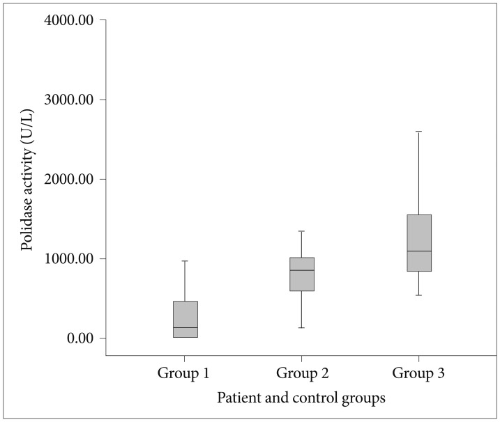
Prolidase activity showed statistically significant differences between the groups when evaluated by ANOVA test (F=14.344, p=0.001). The groups were compared as pairs using the post-hoc Tamhane test. Prolidase activity showed statistically significant differences between the patient and control groups, and also between the two control groups [p=0.045 (group 1–2); p<0.001 (group 1–3); and p=0.006 (group 2–3), respectively]. Prolidase activities in the patient and control groups are illustrated in Figure 1.

Prolidase activities in the patient and control groups. Group 1: patient group, Group 2: individuals experienced the earthquake but did not exhibit PTSD symptoms group, Group 3: healthy group. PTSD: post traumatic stress disorder.
PCL-C and CGI scores in group 1 were 48.80±16.31 and 4.00±0.61, respectively. In group 2, PCL-C scores was 23.35±5.34. The differences in PCL-C scores between the groups were found to be statistically significant (t=7.549 p<0.001).
The correlations between the prolidase activity and age, educational level, and BMI were evaluated in the patient and control groups. There was no correlation between the prolidase activity and age, educational level, or BMI in the patient and control groups (p>0.05).
The correlations between the prolidase activity, and PCL-C and CGI scores in group 1 were evaluated. No correlation was found between the prolidase activity and PCL-C or CGI scores in group 1 (p>0.05). The correlations between the prolidase activity, and PCL-C scores in group 2 were evaluated. No correlation was found between the prolidase activity and PCL-C scores in group 2 (p>0.05).
The correlation between the prolidase activity and smoking status was also evaluated. Prolidase activity did not differ significantly between smokers and non-smokers in the patient and control groups (p>0.05).
DISCUSSION
The present study is the first to evaluate prolidase activity in patients with PTSD. The most important finding of the present study is the significantly low prolidase activity in the Patient Group when compared to the control groups. In addition, prolidase activity was found to be significantly lower in individuals experienced the earthquake but did not exhibit PTSD symptoms groupcompared to healthy group.
The studies have reported a decrease in prefrontal cortex activity and the observation of structural changes in hippocampus in patients with PTSD.3132 The studies have also reported an increased production of free radicals as a stress response in the hippocampus, and increased oxidative stress was suggested to mediate hippocampal damage.3334 Nitric oxide (NO) is an oxidative molecule, and has been suggested to exert its excitatory effects on the central nervous system by activating guanylate cyclase in the presynaptic terminal. Hence, causing the release of glutamate via cGMP and increased glutamate NMDA receptor activation in the postsynaptic membrane.353637
An excessive glutamate release in the hippocampus and prefrontal cortex during traumatic events causes oxidative stress and excitotoxicity. The activation of NO, glutamate, and NMDA receptors have major roles in stress-related hippocampal degeneration.12383940 It was suggested that sustained NO release in PTSD might be related to neural degeneration. In addition, hippocampal atrophy and cognitive deficits associated with PTSD have been related to increased release of NO and glutamate and hyperactivation of NMDA receptors.4142434445 NMDA and Nitric Oxide Synthase (NOS) antagonists were shown to be effective in the treatment of PTSD. Furthermore, antidepressants used in the treatment of PTSD were suggested to decrease glutamate level and inhibit NOS.43464748495051
Proline, a non-essential amino acid, is synthesized from glutamate by the reversal of reactions in proline catabolism.52 Endogenous extracellular proline is considered to increase the effects of glutamate in the synaptic cleft.53 Increased proline levels were shown to activate NMDA receptors.53545556 Prolidase has an important role in the regulation of NO biosynthesis. NO, which is involved in many biological processes, and prolidase are considered to have a strong relation with each other.575859 NO is shown to increase in cells in repairement process released by macrophage cells that are activated as part of the immune response. 5960 It is shown that NO plays an important role in collagen metabolism.59 Because of the fact that approximately %25 of the amino acids in the collagen tissue consist of proline and hydroxyproline, prolidase plays an important role in collagen metabolism.61 It was shown that Nitric Oxide regulates prolidase activity with phosporylation of prolidase enzyme through cGMP kinase.59
Glutamate and NO levels are elevated in PTSD and have been documented for their neurotoxic effects.45 The decreased prolidase activity in PTSD may cause a decrease in NO and glutamate levels. This notion has been supported by the regulatory role of prolidase in the synthesis of NO. Proline is excreted in the urine when prolidase activity decreases.62 Proline is synthesized from glutamate.52 The decrease in proline level can be restored from glutamate, and this can also decrease glutamate levels. In addition, decreased proline levels can lead to a decrease in glutamatergic activity in the synaptic cleft and inhibition of NMDA receptors. These mechanisms have led to our suggestion that decreased prolidase activity might produce a neuroprotective effect that prevents neurotoxic effects of NO and glutamate.
Lower prolidase activity in individuals experienced the earthquake but did not exhibit PTSD symptoms group when compared to healthy group (and lower prolidase level in patients when compared to individuals experienced the earthquake but did not exhibit PTSD symptoms group) also lead to the consideration that prolidase plays a compensatory role in the neurotoxicity increased by stress.
Increasing prolidase activity was shown to be related with oxidative-stress in bipolar disorder.20 However it does not seem possible to establish a relationship between prolidase activity that decreases in PTSD and oxidative-stress due to the fact that oxidative-stress levels in PTSD are generally similar to that of healthy individuals.6364 However it was found that NO levels are increased in bipolar disorder like in PTSD.6566 It is found that prolidase activity is increased in schizophrenia patients according to healthy controls.67 It was suggested that Glutamatergic/NMDA receptor dysfunction could play a role in the potential pathogenesis of the schizophrenia patient.6869 Just as increasing prolidase activity in schizophrenia and bipolar disorder may cause NO, glutamate and NMDA receptors' dysfunction, resulting in being associated with neurodegeneration, decreasing prolidase activity in PTSD could thus be associated with neuroprotective effect.
No difference was found between the patient and control groups in terms of gender, age, educational level, BMI, and smoking status. While creating the study groups, subjects in the patient and control groups were selected from individuals that had similar sociodemographic characteristics. The present study showed in whom trauma could influence prolidase activity independent from other factors.
In previous studies, prolidase activity was found to be related with oxidative stress and NO which is an oxygen radicale. The relationship between these parameters and the prolidase activity has been evaluated since oxidative stress can be affected by age, gender, smoking and body mass index (BMI).2022242559 In the patient and control groups, no correlation was found between prolidase activity and age, gender, BMI, smoking status. A study by Yıldız et al. evaluated prolidase activity in patients with coronary artery disease and found no relation between smoking status and prolidase activity. This finding supports and is consistent with our current results.70 We put forth the notion that neural damage associated with smoking may be related to the number of cigarettes smoked, duration of smoking, and age smoking onset. The above-mentioned factors were not investigated in detail in the present study. Therefore, it is not possible to draw conclusions about the effects of smoking on prolidase activity.
Limitations of this study include low patient numbers of study groups and non-evaluation of NO and glutamate levels along with prolidase activities, single measurement of prolidase activity and non-evaluation of post-treatment prolidase activity.
We suggest that the decrease in prolidase activity in PTSD could result in a neuroprotective effect. However, repetitive measurements where NO and glutamate levels are evaluated along with prolidase activity is necessary in order to have a clear identification of the neuroprotective effect of decrease in prolidase activity in PTSD with wider patient series.Molecules that affect prolidase activity must be considered when determining treatment options for PTSD.

