INTRODUCTION
Generalized anxiety disorder (GAD) affects an average of 5% of people over the course of their lives and up to 25% of patients who visited anxiety disorder clinics have GAD.1 GAD is characterized by chronic, excessive and difficult to control anxiety and worry that is associated with somatic symptoms,1 which might be due to abnormalities of the functions that regulate emotional processes.2 Normally, people tend to use worry as a strategy for managing emotional self-control. However, excessive worry is a fallacious strategy to solve objective and subjective difficulties, which cause uncontrollable anxiety, and vice versa. Cognitive models suggest that worry reflects an overlearned compensatory strategy for dulling emotional experience.3
Despite a growing recognition of the importance of emotion regulation deficits in GAD, few studies have assessed neural mechanisms of the effects of emotion on cognition. To our best knowledge, this study is the first fMRI study examining the regional brain differences from the direct effects of emotional distraction during delayed working memory (WM) task among GAD patients. We evaluated the effects of emotional distractors on WM maintenance in GAD patients using functional Magnetic Resonance Imaging (fMRI) with a face recognition task.
METHODS
Subjects
Fifteen GAD patients (mean age=36.4┬▒11.2 years) participated. Written informed consent was obtained from all participants before participation. All of the GAD patients were diagnosed on the basis of DSM-IV-TR by using Structured Clinical Interview for Axis I DSM-IV Disorders (SCID-I)4 and had no other psychiatric disorders. The mean year of education was 13.7┬▒2.6 years. Except for one patient, psychotropic medications were prescribed for fourteen patients; escitalopram (n=8), paroxetine (n=2), bupropion (n=1), fluvoxamine (n=1), duloxetine (n=1), mirtazapine (n=1), buspirone (n=6), alprazolam (n=5), zolpidem (n=1). Among them, nine patients prescribed multiple psychotropic medication and five patients prescribed single psychotropic medication. This study was reviewed and approved by the institutional review board at the Ethics Committee of Chonbuk National University Hospital.
Task paradigms
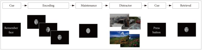
Subjects underwent fMRI scanning during a face recognition task with emotional distractors (neutral and anxiety-provoking pictures). The activation paradigm consisted of a trial with the sequence "encoding (6 seconds); WM maintenance (4 seconds); distracter (6 seconds); button preparation (2 seconds); retrieval (2 seconds); and intertrial interval (ITI) (12 seconds)" (Figure 1). The human faces (half male, half female) were selected from a high school yearbook, converted to black-and-white and cropped into an oval shape showing only the eyes, nose, mouth, and eyebrows. To induce emotional responses of participants, neutral and anxiety-inducing images were viewed. The retrieval task consisted of 10 trials with anxiety-provoking distracters and 10 trials with neutral distracters, yielding a total trial time of 640 s. Each distracter (anxiety-provoking or neutral) trial included two distracter pictures. Of the total 20 trials, the order of the two types of the distracters was randomly arranged. Prior to the fMRI experiment, 50 images of each type were collected from the International affective picture system (IAPS)5 and a variety of Web sites. The neutral images designed to induce a comfortable feeling including scenic pictures and the anxiety-provoking images included photographs of life-threatening behaviors. Ten college students each nominated 30 neutral and anxiety-inducing images from a pool of 100 images as appropriate experimental stimulators. Among them, psychiatrist selected 20 neutral and 20 anxiety-provoking images. All task paradigms for this fMRI study were presented to the subjects using the SuperLab software (Cedrus Corporation, San Pedro, CA, USA).
fMRI data acquisition
An fMRI data acquisition was performed on a 3.0 Tesla Magnetom Verio MR Scanner (Siemens Medial Solutions, Malvern, PA, USA) with a 12-channel bird-cage head coil. A total of 25 axial slices parallel to the anterior commissure to posterior commissure (AC-PC) line were acquired using a gradient-echo echo planar pulse sequence with the following parameters: repetition time (TR)/echo time (TE)=2000 ms/30 ms, flip angle=90┬░, field of view (FOV)=22├Ś22 cm, matrix size=64├Ś64, and slice thickness=5 mm. In addition, two phases of dummy scans were supplemented to circumvent unstable fMRI signals. The high resolution T1-weighted images (TR/TE=1900 ms/2.35 ms) were comprised with FOV=22├Ś22 cm, matrix size=256├Ś256, slice thickness=5 mm.
Data preprocessing and analysis
The fMRI data were analyzed using Statistical Parametric Mapping (SPM8, Wellcome Department of Cognitive Neurology, University College London, London, UK). Prior to the statistical analysis, a slice-timing was performed to correct for differences of the slice-acquisition time. The images were realigned utilizing to the reference volume and were spatially normalized to the standard EPI template in MNI space and resampled to 2├Ś2├Ś2 mm resolution. Finally, the images were smoothed with an 8 mm full-width-half-maximum (FWHM) Gaussian filter. The preprocessed data were analyzed using the standard general linear model (GLM) approach within the SPM8. For the imaging analysis in this study, we used the data of the distracter phase from among the task paradigms. To analyze the individual blood-oxygen-level-dependent (BOLD) signal, an independent t-test was performed in the rest and activation conditions (neutral pictures and anxiety-provoking pictures). For the within group comparison of the GAD patients, the differential brain activation maps, which are correspondent to the contrast of neutral pictures vs. anxiety-provoking pictures, were obtained from the paired t-test (p<0.001) with a spatial extent of at least 10 adjacent voxels.
RESULTS
The demographic characteristics were as follows; age (36.4┬▒11.2), gender (8 male, 7 female), years of education (13.7┬▒2.6). The clinical characteristics were as follows; duration of illness (4.7┬▒7.6), HAM-A (18.5┬▒4.7), STAI-S (53.9┬▒10.5), and ASI-R (62.3┬▒27.1). The feeling of discomfort, which rated after the fMRI experiment, for anxiety-provoking pictures measured on an 11 point visual analogue scale and was significantly higher than that regarding the neutral pictures (7.4┬▒1.5 vs. 0.1┬▒0.4, respectively). In terms of behavioral performance, the scores for the face recognition task with the neutral scene were significantly higher than those for the anxiety-provoking pictures (10 trials each, 72.0┬▒13.2% vs. 60.7┬▒14.9%, p<0.05). There was no missing response in behavioral performance.
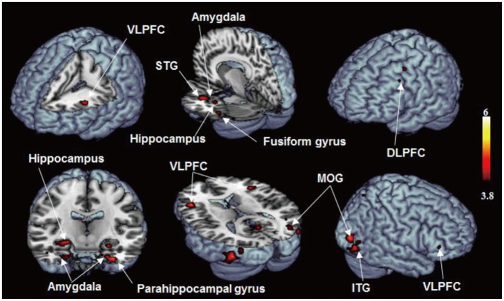
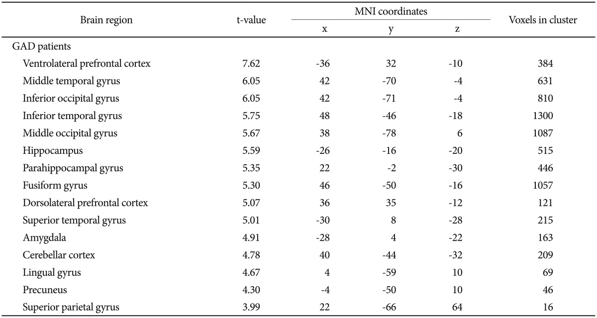
The brain activation area which shown significantly increased activation regarding the anxiety-provoking pictures compared with the neutral pictures were as below (all brain regions p<0.001) (Table 1, Figure 2); VLPFC, Middle temporal gyrus, Inferior occipital gyrus, Inferior temporal gyrus, Middle occipital gyrus, Hippocampus, Parahippocampal gyrus, Fusiform gyrus, DLPFC, Superior temporal gyrus, Amygdala, Cerebellar cortex, Lingual gyrus, Precuneus, Superior parietal gyrus. The brain areas with predominant activities in patients with GAD were not observed when viewing neutral distracter compared with anxiety-provoking distracter (p<0.001).
DISCUSSION
In the present study, we investigated brain activation patterns associated with the effects of emotional distractors during WM maintenance in patients with generalized anxiety disorder. At a facial recognition task level, we found that WM maintenance in the GAD patients was significantly impaired by the emotional distractors, as evidenced by the significantly lower task accuracies with the emotional distracters compared to those with neutral pictures. It seemed that GAD patients tried to maintain goal-relevant in formation in mind and keeping goal-irrelevant information out of mind while interfered by anxiety-provoking situation. Accordingly, in our result, brain regions relevant to anxiety, WM maintenance, and cognitive inhibition showed increased activation. As compared to the face recognition task with neutral pictures, brain regions including the DLPFC, VLPFC, amygdala, and hippocampus revealed increased activation during the task with anxiety-provoking pictures.
Our results are in consistent with previous studies. The role of the amygdala in emotional processing is relatively well established in healthy control67 and in patients with anxiety disorder.8 Amygdala showed greater activity in amygdala in response to fearful faces in GAD patients.910 The hippocampus adjacent to the amygdala also plays a crucial role in anxiety.11 In addition, hippocampal hyperactivity is also associated cognitive dysfunction.12 The prefrontal cortex is highly interacts with various brain structures, including other cortical, subcortical and brain stem sites.13 In healthy controls, the DLPFC involved in the active maintenance of goal-relevant information in WM,1415 whereas the VLPFC was involved in emotional processing.1617 In previous study with GAD patients, Monk et al.18 demonstrated results which shown greater activation in the VLPFC in the attentional bias away from angry face than healthy controls.
Interestingly, it has been suggested that an affective-cognitive interaction mainly constituted by two control system, which are dorsal executive and ventral emotional control system. The dorsal executive control system (such as DLPFC and lateral parietal cortex) involved in the active maintenance of gold-relevant information processing in working memory.141920 The ventral emotional system (VLPFC and medial PFC, and amygdala) involved in emotional processing.202122 Although our results are not conclusive, one possible explanation of increased activation in the DLPFC, VLPFC, and amygdala might be the result of an affective-cognitive interaction during WM maintenance with emotional distracters in GAD patients.
In our result, increased brain activations were found in the fusiform gyrus and superior parietal gyrus. These areas are known to be involved in facial recognition, which also shown increased activation using facial recognition task in previous studies.232425 The inferior and middle temporal gyrus also works together with the fusiform gyrus in recognition of the information.27 The superior temporal gyrus has been involved in the perception of emotions in facial stimuli.28
There are some limitations in our study. First, the number of trials in the WM task used in our study was limited since the head-movement could not be sustained with increasing experimental time during the fMRI. Second, the possibility of medication effects on brain activation patterns could not be excluded.
In summary, the present study provides the first functional neuroimaging evidence in GAD patients that impaired performance in the presence of emotional distracters are associated with brain regions responsible for the active maintenance of goal relevant information in the WM (DLPFC) and emotional processing including the VLPFC, amygdala, and hippocampus. Although our results are not conclusive, our finding cautiously suggests the cognitive-affective interaction in GAD patients which shown interfering effect of emotional distracters on WM maintenance. Further research on the effects of emotion on cognition, such as a comparison with healthy control, may help to further clarify the neural mechanisms of GAD and other anxiety disorders.